RedSafe™ Nucleic Acid Staining Solution
RedSafe™ Nucleic Acid Staining Solution is a new and safe nucleic acid staining reagent, a substitute for EtBr (ethidium bromide).
• RedSafe™ Nucleic Acid Staining Solution is easy and convenient to use, offering higher sensitivity than EtBr.
• It has significantly lower genotoxicity compared to EtBr, ensuring user and laboratory safety.
• When bound to nucleic acids, it becomes visible at 520–540 nm (optimal with a 530 or 535 nm filter).
• Visualization of DNA and RNA bands during agarose gel electrophoresis.
• Isolation of DNA fragments for subcloning while minimizing mutation risks associated with EtBr.
• Both Precasting and Post-Staining are all possible.
| No. | Kit Contents | Unit |
|---|---|---|
| 1 | Staining Solution | 1 ml x 1 vial |
| 2 | Manual | 1 ea |
• RT, Room Temperature : Stable for more than 12 months
• For more stable use, should be store at 4℃ (Stable for more than 24months).
1. Spectrum
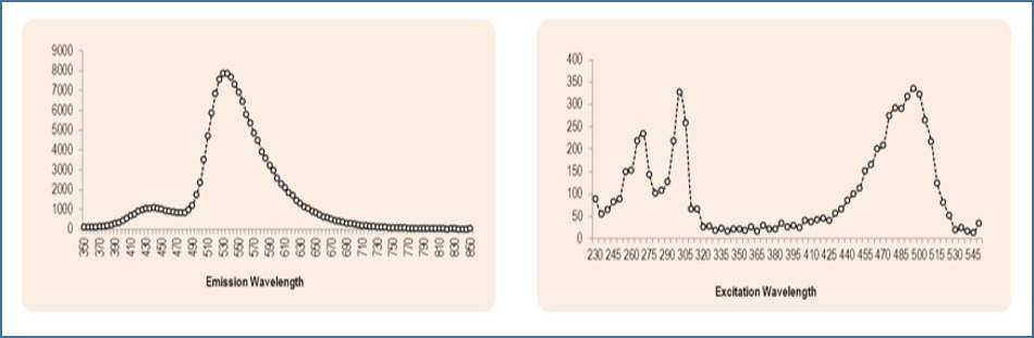
[Excitation (EX) and Emission Wavelengths]
• EM scan at 306 nm EX showed the highest center peak at 530-535 nm.
• EX scan at 535 nm EM revealed multiple peaks, with the center peaks at 270 nm, 300 nm, 480 nm, and 495 nm.
[Excitation Capabilities]
• RedSafe can be excited not only by the UV light typically generated by a UV Transilluminator, but also by a wide range of visible light wavelengths, specifically in the 420-510 nm range.
[Emission Characteristics]
• Upon excitation, RedSafe emits a strong fluorescent signal at 535 nm, which is in the green-yellow region of the visible spectrum.
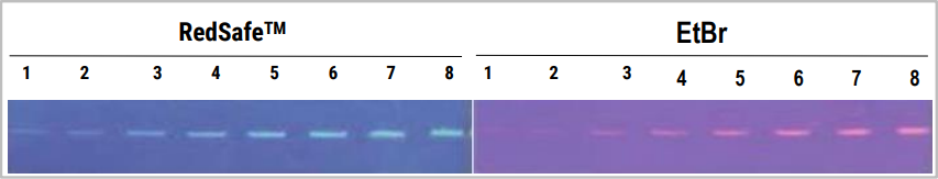
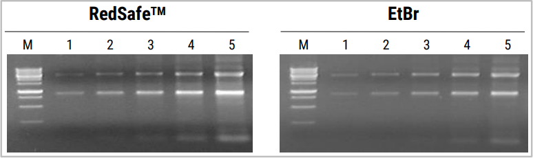
2. Sensitivity
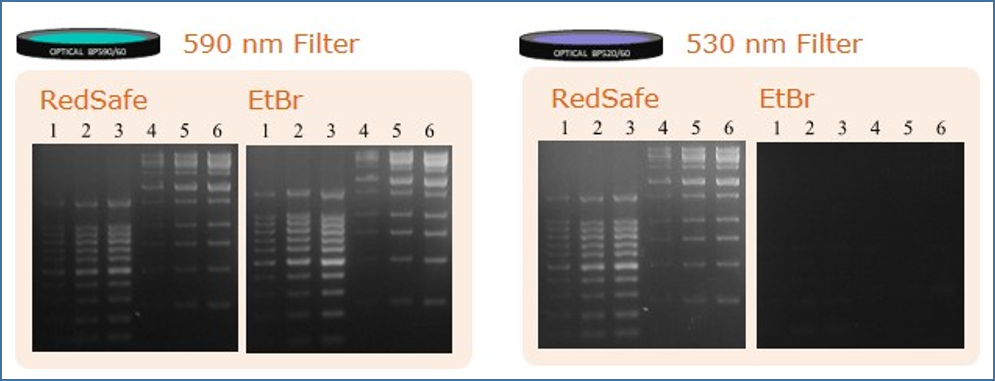
3. Filter Selection
• If using EtBr filter : If the existing laboratory equipment is equipped with an optimal 590 nm filter for EtBr, the fluorescent signal of RedSafe may still be detected, but it may be difficult to obtain the best image.
• If using 520~540 nm filter : When using a filter in the 520-540 nm range, which is suitable for RedSafe, the RedSafe bands will appear brighter and more distinct. If you want to obtain more optimal electrophoresis results using RedSafe, it is recommended to use a 530 or 535 nm filter.
| Product | Cat.No | Capacity | inquire | |||||||
|---|---|---|---|---|---|---|---|---|---|---|
| Maxime™ PCR PreMix (i-Taq) Best |
|
|||||||||
| 2X PCR Master mix Solution (i-Taq) Best |
|
|||||||||
| i-Taq™ DNA Polymerase |
|
|||||||||